Dilated Cardiomyopathy
Dilated Cardiomyopathy initially starts with the degeneration of the heart muscle which becomes weaker and thinner. The heart, especially on the left ventricle should have a thick muscular wall, but over time this muscle becomes thinner leaving less wall and leave more space within the heart. As this happens, the heart loses its ability to pump blood around the dog’s body. The blood congests in the heart, which causes the heart muscle to stretch even thinner and this stretching causes the large, dilated appearance which is where its name comes from. Some dogs may develop a heart murmur if the heart valves become stretched enough to cause a leak. The stretched heart muscle may cause some dogs to develop an unusual heart rhythm.
According to the VCA animal hospitals (https://vcahospitals.com/know-your-pet/dilated-cardiomyopathy-dcm-in-dogs--indepth) “Dilated cardiomyopathy is the most common cause of heart failure in certain large breed dogs. These include boxer dogs, Doberman Pinchers, Great Danes, Irish Wolfhounds, Newfoundland, standard and giant Schnauzers and Saint Bernards.”
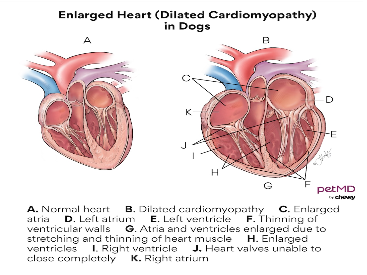
If we look at heart A, in the picture above we can see a normal, healthy dog’s heart. This heart has 4 chambers arranged in a square shape, the bottom 2 chambers are the left and right ventricle and the top 2 chambers are the left and right Atrium. The dog’s blood travels into the right side of the heart, it then moves to the lungs through the pulmonary arteries, in the lungs it picks up oxygen and brings the oxygenated blood back to the dogs heart through the left side of the heart, where the heart then pumps this blood to the rest of the dogs body.
This oxygenated blood fuels the whole of the dog’s body, its cells, its muscles, it helps the body digest the food and with the absorption of nutrients. It also has a role to play in the liver and kidneys being able to get rid of waste products and toxins from the blood. Every single organ in the body needs oxygenated blood to function. This process is essential for life function of the dogs life.
If we look at heart B, in the picture above, we can see a very enlarged heart, you can see the enlarged Atria, the thinning of the ventricle walls, the Atria and Ventricles are enlarged due to the thinning and stretching of the heart muscle. You can also see that the heart valves are not able to close properly. As DCM occurs slowly, over time With heart B not being able to function as it should, if left unnoticed (it is also often missed by many pet owners), it will not be picked up until the dog has gone into (CHF) Congested Heart Failure and often by this point it is FATAL!
Symptoms of DCM in Dogs
As DCM can occur very quickly, it can occur as the blood is not oxygenated enough for the dogs body to function or the blood can back up in the heart making it very difficult for the dogs lungs to function properly, these are just some of the symptoms to watch out for:
- Coughing or gagging
- increased effort when breathing
- rapid breathing when sleeping or resting (more than 30 – 35 beats per minute)
- weakness
- fainting
- restless sleeping, having to change positions
- collapse
- decrease in appetite
- reduced ability to exercise
- quiet and not interacting, depressed
- swollen belly
- weight loss
- Panting
- laboured breathing
- irregular weak pulse
- heart murmur
- crackling or muffled sound when breathing
- sudden death
Although any breed of dog can have DCM, it is much more common to see this appear in dogs aged 4 to 10 years old. While DCM dos not have a definitive cause, it can be hereditary, it has also been proven, to be caused by nutritional deficiencies like a lack of carnitine and taurine in the dogs diet. It can also be caused by infections.
“Dilated cardiomyopathy can occur due to a primary abnormality of the heart muscle itself, but can also be seen secondary to diet, abnormal heart rhythms, infectious causes or metabolic diseases. The overall effect of DCM is a decrease in the contractility (pumping ability) of the heart.”
If you have a breed of dog that is predisposed to DCM then it is in your dogs interests to rotate food regularly, changing between different types of food with different primary ingredients. Foods which are grain free or with primary ingredients of lentils, potatoes, peas or legume seeds have been linked to these conditions. The signs that your dog has DCM is difficult to diagnose especially in the early stages as symptoms may not appear, until sadly it has progressed too far.
How is DCM Diagnosed?
To be able to diagnose that a dog has DCM fully your vet, or heart specialist must perform several different tests on how the different parts of the heart are functioning.
Auscultation: This is done by listening to the dogs heart through a stethoscope, for your vet to ascertain whether the heart valves are closing fully and to listen for a heart murmur. If they hear a heart murmur then they must find the murmurs location and determine its significance. Heart rhythm can also be assessed through auscultation. If there are concerns at this point then your vet or specialist can simultaneously palpate or feel the pulse to determine its rhythm and strength. Auscultation can also be to evaluate the lungs. From experience I have found that most vets and heart specialists cannot evaluate all this by stethoscope, especial on Saints Bernards and Newfoundlands who have a double coats and many other factors influencing the dogs behaviour when visiting a Veterinary practise. If the above valuations cannot be done then further investigation need to be done.
Blood and Urine Tests: These 2 tests are done to screen for underlying kidney disease. Quite often kidney and heart disease are related because the internal organs are often impaired by the lack of oxygen being pumped by the heart. The blood test will look for Cardiac biomarkers including Cardiac Troponin I and ProBNP: these will measure the protein levels in the body that change with structural changes of the heart and when heart disease is present. The vet will analyse these results within the context of the dogs overall condition.
Chest Radiographs – X-rays: A chest x-ray is needed for the vet to examine and assess the lungs. This xray also the allows the vet to examine the shape and size of the heart. DCM can normally be seen as an enlargement of the heart, particularly on the left hand side.
Electrocardiogram (ECG): This assessment allows the vet to measure the electrical activity of the heart. It also allows the vet to be able to accurately measure the heart rate and diagnose any diagnose any abnormal rhythms. If needed a dog can wear a portable heart monitor (portable ECG) for 24 or 48 hours to better assess abnormal heart beats (arrhythmias).
Ultrasound examination (echocardiogram): This procedure is the most accurate to determine the size of the heart chambers and the thickness of the heart walls. By measuring the heart contractions, a vet or heart specialist can accurately evaluate the hearts' ability to pump blood efficiently. A combination of the above test will give the vet a better evaluation of how the dogs heart is functioning. If an accurate diagnosis is made and the severity of the disease ascertained then the proper treatment can be started, if treatment is available. Treating this disease without proper diagnosis could be fatal. If DCM is diagnosed, a referral to a veterinary heart specialist is advisable.
Treatment for DCM
DCM if treatable is most often treated with medicines to decrease the workload of the heart and to remove liquid from the lungs, to make breathing easier and to improve the efficiency of the hearts work. This disease is a managed disease rather than cured. While there is no cure for DCM, those dogs that acquired nutritionally acquired DCM, if caught early, can restore a normal heart function with the correct therapy. Different heart medications are available for DCM, they are:
Pimobendan: this medication lowers the pressure in the veins and arteries and improves the heart muscle strength, this helps to increase the blood flow to the body. Pimobendan is not believed to induce arrhythmias.
(ACE) Angiotensin converting enzyme inhibitors: ACE inhibitors work by lowering blood pressure, reducing the workload of the heart, they also reduce resistance so that it is much easier for the heart to pump the blood back into the body.
Cardiac glycosides: These medications improve the heart function in a number of ways. They slow the heart rate and strengthen the heart contractions, so that the blood is pumped more effectively. Digoxin is the most common cardiac glycoside used by many vets. This medicine though needs to be carefully monitored through routine blood tests and ECG analysis for toxic side effects. This drug has mainly been replaced by Pimobendan except for dogs with atrial fibrillation.
Anti-arrhythmic medicines: Many dog with DCM also experience arrhythmias. These medicines can be used alongside the above drugs. These medicines are Beta-blockers and calcium-channel blockers. The drugs works on the hearts electrical system and can only be prescribed by a heart specialist if irregular heart rhythms have been diagnosed.
Nutritional Therapy: Dietary changes and nutritional support may be needed. These may include taurine and carnitine supplementation. There is a ongoing study to see if dogs with diet induced DCM can return to normal with diet changes alone. FDA Investigation
Bronchodilators and cough suppressant medication: These medications may be prescribe to make breathing easier and reduce the amount of coughing a dog does with congestive heart failure.
The VCA animal hospital states that; “DCM is a serious disease that must be accurately diagnosed and aggressively treated.”
Sadly there is no magic cure for a dog who is diagnosed with DCM. It will all depend on the dogs diagnosis at the time. Age, health, how the heart is actually functioning, For some dogs medication can slow down the progression for others if DCM has progressed, this will make their prognosis worse. Some dogs will do well with treatment , some will be able to resume a normal life. For those dogs who have DCM diagnosed at a very late stage, the survival rate is between 2 and 24 months.
If you are interested in learning more about DCM and CHF please see the research papers below.
https://www.sciencedirect.com/science/article/abs/pii/S1760273421001077
https://www.sciencedirect.com/science/article/abs/pii/S1760273421001004
https://onlinelibrary.wiley.com/doi/full/10.1111/jvim.15972
https://onlinelibrary.wiley.com/doi/abs/10.1111/j.1939-1676.1995.tb03266.x
https://avmajournals.avma.org/view/journals/javma/253/11/javma.253.11.1390.xml
https://onlinelibrary.wiley.com/doi/full/10.1111/jvim.16397
Hip Dysplasia – What is it?
The hip joint operates as a ball and socket joint. In dogs which have hip dysplasia the ball and socket do not fit properly together. Therefore instead of the joint running smoothly, it will often grind and rub bone against bone, or bone against cartilage. If this is left untreated it, the joint will deteriorate resulting in the loss of operation of the hip joint altogether.
Hip Dysplasia can occur when a dogs hip joints develop incorrectly, allowing the femoral head to partially or fully dislocate.
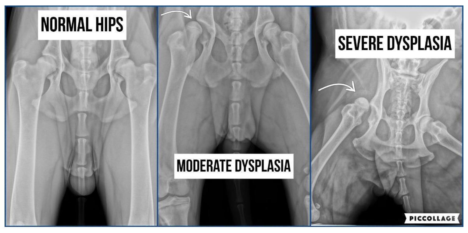
As you can see from the images above (taken from https://pattonvethospital.com/blog/235290-hip-dysplasia-in-dogs) We can see in the first image the normal hips of a dog, where the ball and socket hip joints fit very well together. The hip joint fits snugly and the ball and socket move as they should.
The middle image shows a dog that has moderate hip dysplasia, as you can see the joint of the right of this image fits a little bit high in the joint but the left hip joint is not sitting right at all in that socket. In this image we can see the space in the socket is greater than in the first image. A dog with moderate hip dysplasia will be in moderate pain, with mild lameness when walking.
The image on the rights show a dog that has severe hip dysplasia as you can see both hips are not sitting in any socket whatsoever. The balls and sockets are completely separated. Hips and spine are not straight at all. A dog with this condition may only be able to walk with great difficulty, if at all. He will need surgery.
According to Fitzpatrick referrals “Hip Dysplasia is the commonest orthopaedic condition in dogs. It most frequently affects large rapidly growing dogs.” The signs that your dog has hip dysplasia can be varied but “it is most commonly diagnosed between 6 and 12 months of age.”
Symptoms of Hip Dysplasia in Dogs
- Decrease in activity
- Stiffness
- “Bunny Hopping” gait
- Hip Pain and Sensitivity
- Difficulty standing up
- Difficulty getting in or out a car
- Inability to climb stairs or jump
- Limping on one or both legs
- Protection of hip area when being bathed or groomed
- Weight shifting to front limbs
- Loss of muscle mass in one or both hind legs
How is Hip Dsyplasia diagnosed?
Symptoms are usually seen by an owner in the first instance, a vet will then evaluate the dog and a specialist orthopaedic surgeon will do the operation, depending on the severity of the disease.
If the vet suspects that your dog has hip dysplasia he will need to take x-rays and radiographs to show the severity. The vet or orthopaedic surgeon must carry out a full orthopaedic assessment on the dogs hips and legs. From this they may well be able to give you some results and diagnosis but sometimes more radiographs need to be done under general anaesthetic or sedation, in order gain better specifics of the joints. On top of this an MRI or CT scan may also be necessary.
Treatment of Hip Dysplasia
Depending on the severity of the disease, most vets will recommend;
- Weight control – keep your dogs weight on the leaner side as to not put too much stress on the hip joints
- Rest – dogs with HD will need periods of rest, especially if the disease is causing them discomfort and pain
- Anti-inflammatory pain relief (NSAIDs) or other pain medication prescribed by the vet
- Controlled Exercise – regular short walks on lead are advisable rather than letting your dog off lead to run around madly
- Avoid, racing around, skidding, jumping and long walks
- If your dog does not respond to the above then Surgery may be needed
- There are a few different operations that your vet could offer you, but most operations are carried out by a specialist veterinary hospital
There are some ongoing treatments and care that you can consider if your dog is diagnosed with HD, they are:
- Hydrotherapy – this is a great way to exercise your dog without putting the strain on their joints
- Physiotherapy – carried out by a professional can help build up the dogs muscles which wil take the pressure off the hip joints
- Joint supplements – these may slow down the onslaught of arthritis
Hip Dysplasia is a very painful condition that requires lifelong treatment. Most dogs with HD develop arthritis in the hip joints as they age.
If a dog has mild HD they may live a full and happy life, as they should respond well to controlled daily management or pain relief, exercise and weight control. Many dogs that require surgery can also go on to lead happy, fulfilled lives. For a few dogs, when the pain is severe and also becomes uncontrollable then surgery may not be an option, sadly at this point euthanasia is the only option.
Preventing Hip Dysplasia
Breeders can prevent hereditary HD by screening for this disease before they breed their dogs. There are Kennel Club and BVA (British Veterinary Association) health requirements for each breed of dogs. Before buying a dog, ask the breeder about the hip scores of both parents, also do your research and find out what the average hip score if for that breed of dog. The higher this score the more at risk the dogs are of experiencing HD. The mean score for Saint Bernards in the last 15 years in the UK 21.1. So the breeding parents hip scores should be lower than this.
Although HD is predominately a genetic disease, it can also be affected by diet, exercise, the environment, muscle mass, growth rates and hormones. Why do some puppies develop HD whereas a sibling may not? This could be due to excessive calorie intake which causes rapid growth and weight gain, causing too much stress on the joints. Excessive exercise as a puppy can also cause the onset of HD, due to the ligaments and bones not being fully formed and still growing.
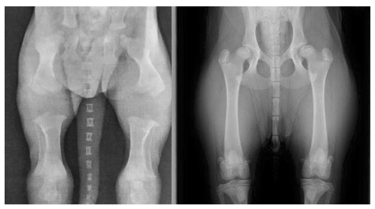
https://freyathecockapoo.com/2017/10/02/freya-the-cockapoos-first-puppy-walk/
The image on the left is from a 2 week old puppy. As you can see their leg joints do not meet their joints. The image on the right is the same dog at 6 months old. For a Saint Bernard this process can take up to 2 years for the bone plates to completely join together, therefore at any time during this stage if the dogs bones are hurt or injured, this could cause hip or elbow dysplasia, it can cause bones to form abnormally, too much exercise or rough play can cause these soft bones to break more easily too, therefore, it is essential to know your breed. Giant breed of dogs often do not reach their full adult size till between 18 months and 2 years.
If you are interested in learning more about hip dysplasia in Dogs these articles, research papers below may help you
https://www.pure.ed.ac.uk/ws/portalfiles/portal/226229318/s12864_021_07945_z.pdf/
https://www.sciencedirect.com/science/article/abs/pii/S0167587700001409/
https://www.frontiersin.org/articles/10.3389/fvets.2019.00490/full/